1) The right ventricle is the chamber of the heart that pumps blood for the pulmonary circulation. Based on this information, blood from the right ventricle is on its way to the __________.
lungs
liver
appendages
2) Which of the following is correct regarding the flow of blood in reference to the left side of the heart?
Blood flows from the left atrium, through the bicuspid valve, into the left ventricle, through the aortic semilunar valve, and then into the aortic arch.
Blood flows from the left atrium, through the aortic semilunar valve, into the left ventricle, through the bicuspid valve, and then into the aortic arch.
Blood flows from the left atrium, through the bicuspid valve, into the left ventricle, into the aortic arch, through the aortic semilunar valve, and then into the systemic arterial system.
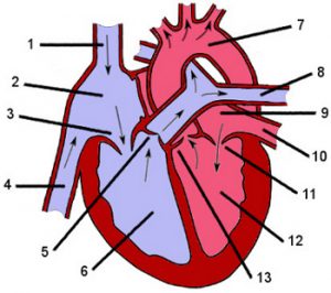
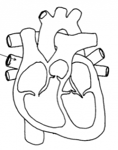
3) Number 2 is pointing to …
the right atrium
the left atrium
the right ventricle
4) The aorta is indicated by number …
The anatomy of the heart
1
8
7
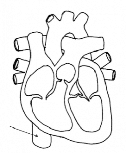
5) Number 1 is pointing to …
The anatomy of the heart
the aorta
the superior vena cava
the inferior vena cava
6) What is number 8 pointing to?
The anatomy of the heart
pulmonary arteries
pulmonary veins
aorta
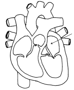
7) Number 4 is pointing to …
The anatomy of the heart
the pulmonary valve
the inferior vena cava
the superior vena cava
8) The pulmonary valve is indicated by number …
The anatomy of the heart
12
5
11
9) The tricuspid valve is indicated by the number …
The anatomy of the heart
3
12
11
10) The inferior vena cava is indicated by number …
The anatomy of the heart
1
5
4
11) The right ventricle is indicated by number …
The anatomy of the heart
6
4
13
12) The distal end of the heart
pericardium
epicardium
apex
myocardium
13) inner layer of the pericardium
parietal pericardium
visceral pericardium
myocardium
endocardium
14) outer layer of the pericardium
visceral pericardium
parietal pericardium
epicardium
myocardium
15) The arteries that feed the heart muscle
Capillaries
Coronary
Chordae teindineae
Coronary sinus
16) After blood leaves the Right atrium it passes through the ________.
right ventricle
aortic valve
av valve
tricuspid valve
17) Blood may exit the heart through either ______ or ______ structures called the great vessels.
superior vena cava, inferior vena cava
pulmonary trunk, aorta
the right ventricle left ventricle
pulmonary arteries, pulmonary veins
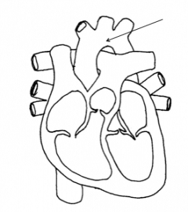
18) Name the part.
Pulmonary vein
Right atrium
Aorta
Pulmonary artery
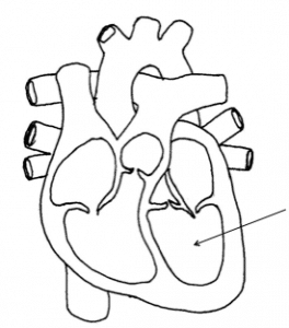
19) Name the part.
Left ventricle
Vena cava
Right atrium
Right ventricle
20) Name this part.
Superior vena cava
Right ventricle
Inferior vena cava
Pulmonary vein
21) Name this part.
Pulmonary veins
Left atrium
Aorta
Pulmonary artery
22) Name this part.
Right atrium
Left ventricle
Right ventricle
Left atrium
23) Which side of the heart pumps oxygen-rich blood to the rest of the body?
Right
Left
24) Which side of the heart pumps blood into the lungs?
Right
Left
25) ______ carry blood to the heart.
Arteries
Veins
Capillaries
Vena Cava
26) _____ carries blood away from the heart.
Blood cells
Veins
Capillaries
Arteries
27) What carries the oxygenated blood from the lungs to the left atrium of the heart?
vena cava
aorta
pulmonary vein
pulmonaryartery
28) What are the two large veins that drain blood from the upper body and from the lower body and empty it into the right atrium of the heart?
vena cava
aorta
pulmonary artery
pulmonary vein
29) The two chambers of the heart with thin walls that collect blood from the major veins and empty it into the larger, more muscular chambers.
aorta
atria
ventricles
lungs
30) What are the functions of the tricuspid and bicuspid valves?
prevent blood from leaking back into he ventricles
prevent blood from leaking back into the atria
allow blood to flow into the atria
allow blood to leave the ventricles
31) What is the main function of the heart?
The heart circulates blood and oxygen throughout the body.
The heart is the control center for all body activities.
The heart gets rid of the wastes in your body produced by homeostasis.
The heart transports, protection, and regulation.
32) What does the left side of the heart do?
The left side of the heart pumps blood through the lungs where it gets oxygen.
The left side of the heart receives the blood containing oxygen and pumps blood to the rest of the body.
The left side of the heart controls the muscles on the left side of the body,
The left side of the heart supply our arms with blood
33) What does the right side of the right heart do?
The right side of the heart receives deoxygenated blood from the body through the vena cava and pumps it into the right ventricle which then sends it to the lungs to be oxygenated.
The right side of the heart is responsible for words, logic, numbers, analysis, lists, linearity and sequence
The right side of the heart pumps blood through the lungs where it gets oxygen.
The right side of the heart is a meaty organ that sits on the right side of the belly.
34) Name part #6
Left Ventricle
Left Pulmonary Artery
Left Pulmonary Vein
Aorta
Heart Anatomy Questions And Answers
Superior Vena Cavae
Right Atrium
Tricuspid Valve (right)
Right Ventricle
Pulmonary Valve
Pulmonary Veins
Left Atrium
Mitral Valve (left)
Left Ventricle
aorta
Pulmonary valve (semilunar valves; DUB)
aortic valve
endocardium
pericardium
interventricular septum
papillary muscles
chordae tendonae
trabeculae carnae
pectinate muscles
moderator bands
fossa ovalis
pulmonary trunk
pulmonary arteries
ligamentum arteriosum
inferior vena cavae
opening of coronary sinus
apex
The anatomy of the heart consists of
Chambers, valves, vessels and circulation
The function of the heart is to
circulate blood throughout the lungs and various tissues of the body.
Without the circulation of the heart
nutrients cannot be delivered, waste products cannot be removed, and therefore you would die.
What happens when the heart contracts
blood moves into the lungs to get oxygen, then the oxygen gets into the body’s circulation delivering oxygen to the tissues
Blood supplies the tissues with
nutrients and O2
Blood removes
CO2 and waste products
The heart is what shape organ
triangular shaped
The heart is located
in the center of the chest, under the sternum and in between the lungs. 2/3 rds of the heart lies to the left of the sternum
The Approx. size of the heart is
The size of a fist
The approx weight of the heart is
10.6 oz
Beats average (minutes, Day, lifetime)
72 beats per minute – 100,000 beats per day – 22.5 billion in a lifetime
The heart Pumps about ____ ml blood per beat for a total ___L/minute
140 blood per beat for a total 5L/minute
Heart pumps daily about ______ L or ______ gallons of blood (average bathtub filled 36 times)
7250 L or 1800 gallons of blood average bathtub filled 36 times
The heart is like ____ pumps side by side divided by ________.
2 pumps side by side divided by septum
What are the 4 chambers of the heart
2 upper atria 2 lower ventricles
What keeps blood flowing in one direction
Valves
What prevent the valves from turning inside out
Chodae Tendae
Middle muscular layer
Myocardium
Muscle is thickest in what chamber of the heart
L ventricle
thicker muscle in heart chamber means.
stronger contraction of that chamber giving it the ability to pump blood through body
The valve between the R atrium and the R ventricle is the
Tricuspid valve
The valve between the L atrium and the L ventricle is the
Bicuspid valve AKA mitral valve
Valves that divide the atrium from the ventricles on R & L of heart are known as
Atrioventricular valves
Why are the heart valves one-way
to keep the blood flowing in one direction
What type of valve is named because they look like half moons. And what are the names of the 2 in your heart
returns blood from the head, arms and upper body to heart
Superior vena cava
returns blood from the lower legs and body to heart
Inferior vena cava
when the heart contracts the R ventricle pumps deoxygenated blood to the lungs via this artery
Pulmonary artery
transports oxygenated blood from the lungs back to the heart via L atrium
Pulmonary veins
The largest artery in the body. It transports oxygenated blood to entire body. t
Aorta
The first of the arteries to branch off of the aorta is
coronary arteries
Trace a drop of blood through the heart to the lungs
Enters via the vena cava to R atrium -Thru the tricuspid valve -Into R ventricle -Thru pulmonary valve -To pulmonary arteries to lungs -There are tiny capillaries and thin walls so O2 and CO2 can pass over through Osmosis
Trace a drop of blood from the lungs through the heart to the body
Blood now has oxygen -Thru pulmonary veins -Into L atrium -Past Mitral (bicuspid ) valve -Into L ventricle -Out the aorta (largest artery in the body) -The aorta after descending past the diaphragm is known as the abdominal aorta
Three types of circulation
Pulmonary – between the heart and lungs-Systemic – between the heart and body-Coronary – the hearts blood supply
The portion of the cardiovascular system which carries oxygenated blood away from the heart, to the body, and returns deoxygenated blood back to the heart.
Systemic circulation
Refers to the movement of blood through the tissues of the heart
Coronary circulation
Each beat of the heart has two phases that indicate contraction and relaxation periods what are the two phases and what cycle do they make up
Relaxation phase – Diastole Conduction Phase – Systole -These 2 phases together make up the cardiac cycle.
What happens during diastole
Blood from the body returns to the heart via vena cavas. R atrium fills with blood and contracts, pushing open the tricuspid valve (this allows blood to flow into the R ventricle). At the same time, blood returns from the lungs via the pulmonary veins into the L atrium . (blood fills L atrium prior to atrial contraction). Atrial contraction forces mitral valve to open to allow blood to flow into L ventricle.
What happens during Systole
Heart muscles contract, creating pressure to open the pulmonary and aortic valves. Blood from R ventricle is pushed into the lungs to exchange O2 and CO2. Blood from the L ventricle is pushed thru the aorta to be distributed throughout the body.
In general women have faster heartbeats then men true or false
true
Children’s rates are faster then adults true or false
true
On average adults heart beats
60-100 bpm
what sound is heard during systolic phase by the contraction of the ventricles and the closing of the AV valves
Lubb sound
during the diastolic phase. It is a shorter sound and occurs during the beginning of ventricular relaxation. Caused by the closing of the semilunar valves.
Dubb sound
Each contraction of heart is caused by
electrical impulses throughout the heart
The pumping cycle of heart is controlled by
electrical impulses
______ _______are initiated and transmitted through the heart in a specific pathway.
Electrical impulses
What do Specialized masses of tissues in the heart do
They produce electrical impulses and form the conduction system
The conduction system is necessary
for the heart to pump continuously and rhythmically.
ability of the heart to initiate an electrical Impulse without being stimulated by another source
Automaticity
Ability of the heart muscle cells receive and transmit an electrical impulse
Conductivity
Ability of the heart muscle cells to shorten in response to an electrical stimulus
Contractility
Ability of the heart muscle cells to respond to an impulse or stimulus. Without this quality the heart would nor react to the electrical impulses that are initiated within the heart
Excitability
Where is the SA node located
in upper portion of the R atrium
What is the SA node also known as? /why?
AKA pacemaker of the heart – it initiates the heartbeat
Automatricity of the fibers of SA node produces
the contraction of the R and L atria
SA node fires about
60 – 100 times per minute
Where is the AV node located
on the floor of the R atrium
Impulses travel from the SA node à
AV node because of conductivity
The AV node itself causes a delay (slowdown) of the impulse two reasons why
1) to allow additional blood to travel from the atrium to the ventricles before they contract (known as atrial kick) this will increase the cardiac output (the amount of blood pumped out of the heart into the rest of the body) 2) reduces the number of electrical impulses transmitted onto the ventricles, this is important if the SA node is firing too fast
What is located next to the AV node – Provides transfer of electrical impulses from atria to the ventricles
Bundle of HIS
What is located on the L and R side of the interventricular septum- Impulses travel R and L bundle branches to the R and L ventricles Electrical impulse split and travel down both sides. This activates the myocardium to contract.
Bundle branches
What Speeds impulses thru the ventricles provides an electrical pathway for each of the cardiac cells. The electrical impulses accelerate and activate the L and R ventricle at the same time to contract.
Purkinje fibers
Was this helpful?
Let us know if this was helpful. That’s the only way we can improve.
The Quizzma Team is a collective of experienced educators, subject matter experts, and content developers dedicated to providing accurate and high-quality educational resources. With a diverse range of expertise across various subjects, the team collaboratively reviews, creates, and publishes content to aid in learning and self-assessment.
Each piece of content undergoes a rigorous review process to ensure accuracy, relevance, and clarity. The Quizzma Team is committed to fostering a conducive learning environment for individuals and continually strives to provide reliable and valuable educational resources on a wide array of topics. Through collaborative effort and a shared passion for education, the Quizzma Team aims to contribute positively to the broader learning community.


")


")


")